
OLIGODENDROCYTE PROGENITOR CELLS (GRNOPC1)
Geron's hESC-Derived Oligodendrocyte Progenitor Cells - GRNOPC1
Geron's first product manufactured from hESCs to enter clinical testing
is GRNOPC1. GRNOPC1 is a population of living cells containing
precursors to oligodendrocytes, otherwise known as oligodendrocyte
progenitor cells (OPC). Oligodendrocytes are naturally occurring cells
in the nervous system that have several functions. Oligodendrocytes
produce myelin (insulating layers of cell membrane) that wraps around
the axons of neurons to enable them to conduct electrical impulses.
Myelin enables efficient conduction of nerve impulses in the same manner
as insulation prevents short circuits in an electrical wire. Without
myelin, many of the nerves in the brain and spinal cord cannot function
properly. Oligodendrocytes also produce neurotrophic factors
(biologicals that enhance neuronal survival and function) to support the
maintenance of nerve cells. Oligodendrocytes are lost in spinal cord
injury, resulting in myelin and neuronal loss that cause paralysis in
many patients with spinal cord injuries.
In preclinical studies, GRNOPC1, when injected into the injury site of
spinal cord-injured animals, migrate throughout the lesion site and
mature into functional oligodendrocytes that remyelinate axons and
produce neurotrophic factors (Stem Cells and Development, Vol. 15, 2006), resulting in improved locomotion in the treated animals.
The ultimate goal for the use of GRNOPC1 in man is to achieve spinal
cord repair by injecting these cells directly into the spinal cord
lesion.
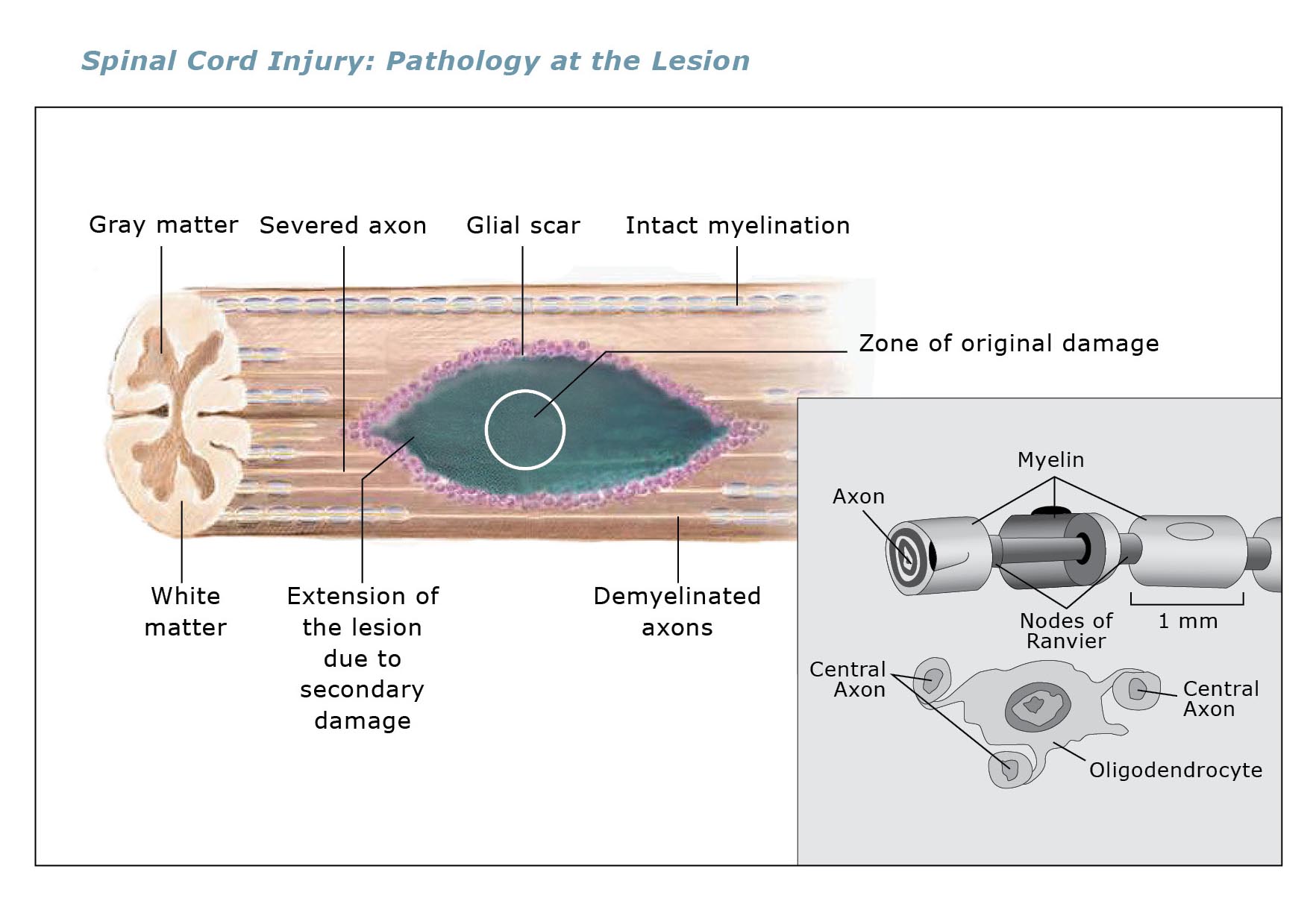
The Pathology of Acute Spinal Cord Injury
Most human spinal cord injuries are due to contusions (bruises), rather
than to a severing of the spinal cord. Most automobile, sports or
industrial accidents cause a displacement or crush of the vertebral
bodies in the cervical or thoracic spine that results in a contusion
within the spinal cord, damaging the delicate nerve fibers at the site
of the fracture. Spinal cord injuries cause severe inflammation within
the spinal cord at the site of the fracture that is particularly toxic
to the oligodendrocytes in the spinal cord.
GRNOPC1 Restores Locomotion in Rodent Models of Spinal Cord Injury
Geron scientists, in collaboration with Dr. Hans Keirstead's laboratory
at the University of California, Irvine, developed a method to produce
oligodendrocyte progenitor cells from hESCs. These cells were tested in a
validated rodent model of acute spinal cord injury. Under anesthesia,
animals were given a spinal cord contusion injury, mimicking what occurs
in humans who suffer traumatic injury to the spinal cord. The lesions
resulted in loss of truncal muscle function, bladder control and hind
limb function. The injured animals received either no treatment, control
cells or media, or one injection of GRNOPC1 within seven days after
injury. Animals were then carefully followed and observed for locomotor
recovery after the injury.
Injured animals treated with GRNOPC1 displayed significant improvement
in a variety of functional parameters compared to control groups.
GRNOPC1-treated animals had improved hind limb locomotor control. Paw
placement, stride length and paw rotation all significantly improved
compared to controls. When the GRNOPC1-treated animals were examined
histologically, increased remyelination of axons in the injury site was
observed compared to that in the control animals. An increased number of
axons were also observed in the vicinity of the injection within the
injury. These animal results were published in the
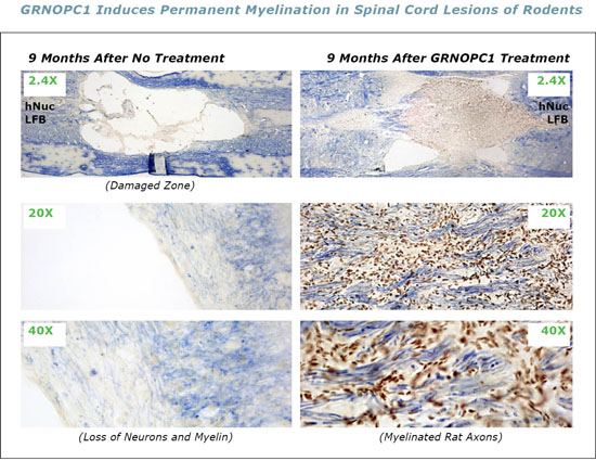
Journal of Neuroscience, Vol. 25, May 2005. In additional
studies, the lesion site of animals nine months after injury and
injection of GRNOPC1 was observed to be essentially filled with GRNOPC1
and myelinated rat axons crossing the lesion. These animal observations
serve as the rationale for the use of GRNOPC1 in treating spinal cord
injuries in man.
Continue to Section 3: Preclinical Safety Studies »
|